-
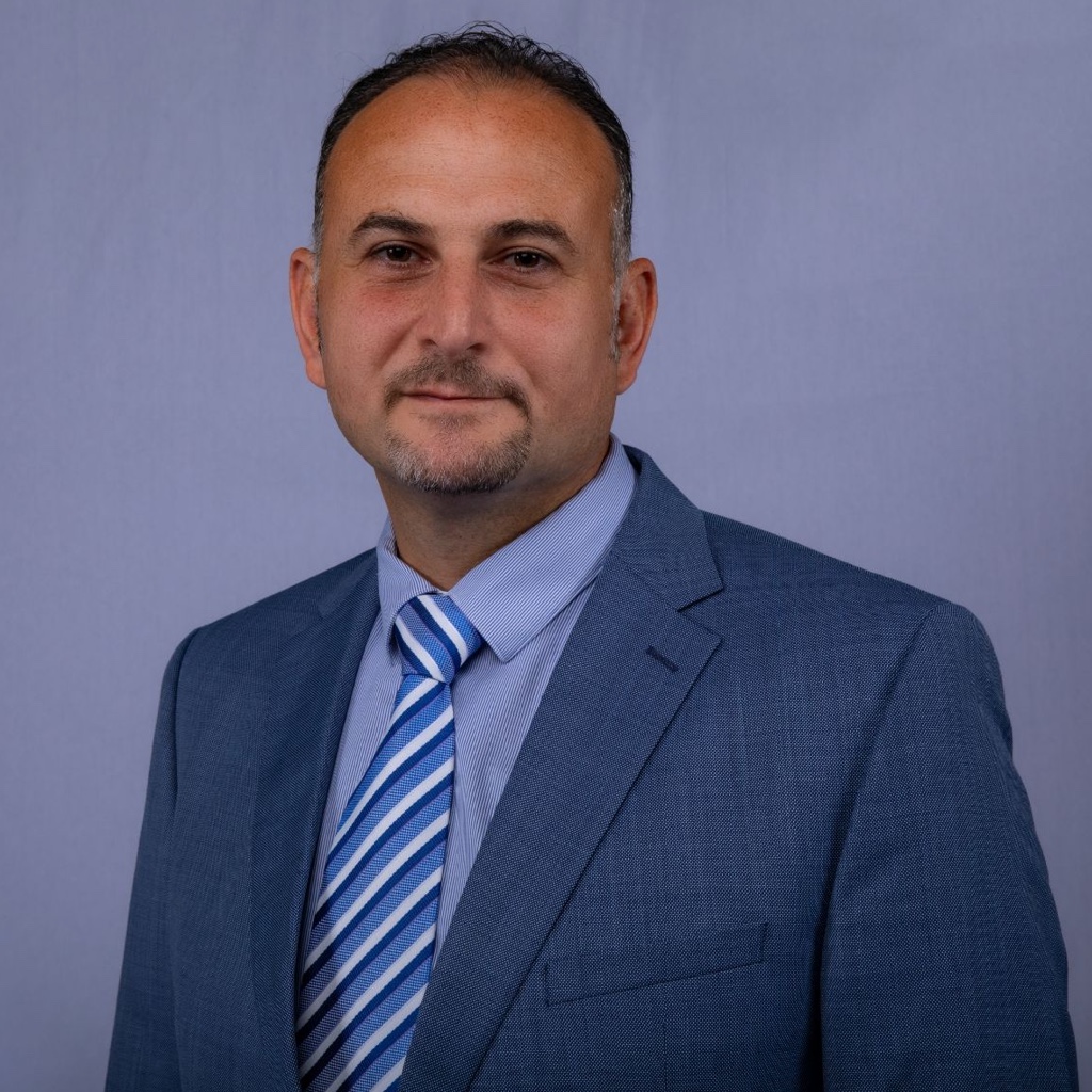
Anas Al-Jadaa
- Associate Professor
- Ext: 2222
Dr. Anas Al Jadaa is an Assistant Professor at the College of Dentistry, Ajman University, since August 2020. He obtained his undergraduate degree in Dentistry from Jordan University of Science and Technology in 2003. With over four years of clinical experience, he advanced his studies at the University of Zurich, Switzerland, where he earned a Doctorate in Endodontics (2010), a Postgraduate Certificate (2011), and a Board Certificate in Endodontics (2012). Dr. Al Jadaa further pursued his academic journey, obtaining a PhD in Clinical Dentistry from the University of Tampere, Finland, in 2016. He is an active member of several esteemed professional organizations, including the Swiss Dental Association, Swiss Society for Endodontology, and the European Society of Endodontics. Throughout his career, Dr. Al Jadaa has supervised numerous master's theses and contributed research to high-ranking dental journals.
Education
- Certificate of Advanced Studies in Periodontology, University of Zurich, Switzerland, 2018
- PhD in Clinical Dentistry, University of Tampere, Finland, 2016
- Swiss Diploma in Dentistry, Ministry of Health, Switzerland, 2013
- Postgraduate Program in Endodontology, University of Zurich, Switzerland, 2011
- Doctorate in Dentistry (Endodontology), University of Zurich, Switzerland, 2010
- Bachelor in Dental Surgery, Jordan University of Science and Technology, Jordan, 2003
Certificate of Advanced Studies in Periodontology, University of Zurich, Switzerland, 2018
PhD in Clinical Dentistry, University of Tampere, Finland, 2016
Swiss Diploma in Dentistry, Ministry of Health, Switzerland, 2013
Postgraduate Program in Endodontology, University of Zurich, Switzerland, 2011
Doctorate in Dentistry (Endodontology), University of Zurich, Switzerland, 2010
Bachelor in Dental Surgery, Jordan University of Science and Technology, Jordan, 2003
Experience
- Associate Professor, Department of Clinical Sciences, Ajman University, United Arab Emirates, Since 2025
- Assistant Professor, Department of Clinical Sciences, Ajman University, United Arab Emirates, Since 2020
- Oberarzt, Department of Conservative Dentistry, University of Zurich, Switzerland, 2017-2019
- Oberassistant, Department of Conservative Dentistry, University of Zurich, Switzerland, 2012-2017
- Wissenschaftlicher Mitarbeiter, Department of Conservative Dentistry, University of Zurich, Switzerland, 2009-2012
- Visiting Doctor, Department of Conservative Dentistry, University of Zurich, Switzerland, 2007-2009
- General Practitioner, Private Clinic, Jordan, 2004-2007
Associate Professor, Department of Clinical Sciences, Ajman University, United Arab Emirates, Since 2025
Assistant Professor, Department of Clinical Sciences, Ajman University, United Arab Emirates, Since 2020
Oberarzt, Department of Conservative Dentistry, University of Zurich, Switzerland, 2017-2019
Oberassistant, Department of Conservative Dentistry, University of Zurich, Switzerland, 2012-2017
Wissenschaftlicher Mitarbeiter, Department of Conservative Dentistry, University of Zurich, Switzerland, 2009-2012
Visiting Doctor, Department of Conservative Dentistry, University of Zurich, Switzerland, 2007-2009
General Practitioner, Private Clinic, Jordan, 2004-2007
Teaching Area
- MSc in Endodontics Clinical and Preclinical
- Advanced Training Program in Endodontics
- Supervision of Master's Theses in MSc in Endodontics
- Supervision of Graduation Projects in the DDS Program
- Clinical Courses in Endodontics within the DDS Program
- Supervision of Postgraduate Doctorate Theses
- Clinical Courses in Preventive Dentistry within the DDS Program
- Dental Morphology Lab
MSc in Endodontics Clinical and Preclinical
Advanced Training Program in Endodontics
Supervision of Master's Theses in MSc in Endodontics
Supervision of Graduation Projects in the DDS Program
Clinical Courses in Endodontics within the DDS Program
Supervision of Postgraduate Doctorate Theses
Clinical Courses in Preventive Dentistry within the DDS Program
Dental Morphology Lab
Research
- Biomechanics of Root Canal Preparation
- Chemo-Mechanical Root Canal Disinfection
- Leakage in Dentistry
Biomechanics of Root Canal Preparation
Chemo-Mechanical Root Canal Disinfection
Leakage in Dentistry
Publications
-
Effect of dentine cutting efficiency on the lateral force created by torque-controlled rotary instruments
-
Influence of the internal anatomy on the leakage of root canals filled with thermoplastic technique
-
Necrotic pulp tissue dissolution by passive ultrasonic irrigation in simulated accessory canals: impact of canal location and angulation
-
Book ChapterAnas Al-Jadaa, Leakage and permeability control in Dentistry: the key for success. Preclinical studies and clinical considerations, PhD dissertation, Leakage and Permeability Control in Dentistry The Key for Success, Tampere University Press, ISBN: 978-952-03-0015-9 (pdf)
-
JournalIbrahim Ali Ahmad 1, Anas Al-Jadaa 2, Three root canals in the mesiobuccal root of maxillary molars: case reports and literature review, Journal of Endodontics, Oct 2014, doi: 10.1016/j.joen.2014.07.034
-
JournalAnas Al-Jadaa 1, Thomas Attin 1, Timo Peltomäki 2, Patrick R Schmidlin 1, Comparison of three in vitro implant leakage testing methods, Clinical Oral Implants Research, Dec 2013, doi: 10.1111/clr.12314
-
JournalAnas Al-Jadaa 1, Thomas Attin 1, Timo Peltomäki 2, Christian Heumann 3, Patrick Roger Schmidlin 1, Impact of Dynamic Loading on the Implant-abutment Interface Using a Gas-enhanced Permeation Test In Vitro, The Open Dentistry Journal, Mar 2015, doi: 10.2174/1874210601509010112
-
JournalF Paqué 1, A Al-Jadaa, A Kfir, Hard-tissue debris accumulation created by conventional rotary versus self-adjusting file instrumentation in mesial root canal systems of mandibular molars, International endodontic Journal, Dec 2011, doi: 10.1111/j.1365-2591.2011.01991.x
-
JournalAnas Al-Jadaa 1, Frank Paqué, Thomas Attin, Matthias Zehnder, Acoustic hypochlorite activation in simulated curved canals, Journal of Endodontics, Oct 2009, doi: 10.1016/j.joen.2009.07.007
-
JournalZainab Shirazi, Anas Al-Jadaa, Abdul Rahman Saleh, Electronic apex locators and their implications in contemporary clinical practice: a review, Open Dentistry Journal, Feb 2023, doi: 10.2174/18742106-v16-e221227-2022-67
-
JournalAnas Al-Jadaa, Zehraa Saidi, Maise Mahmoud, Rahaf Al-Taweel, Matthias Zehnder, Assessment of irrigant agitation devices in simulated closed and open root canal systems, Journal of Endodontics, Apr 2023, doi: 10.1016/j.joen.2023.01.002
-
JournalAnju Daniel, Abdul Rahman Saleh, Anas Al-Jadaa, Waad Kheder, Impact of access cavity design on fracture resistance of endodontically treated maxillary first premolar: In vitro, Brazilian Dental Journal, Mar 2024, doi: 10.1590/0103-6440202405676
-
JournalButhaina Jrab, Abdul Rahman Saleh, Anas Al-Jadaa, Carlos A Jurado, Musab H Saeed, Kelvin I Afrashtehfar, Fracture resistance of CAD/CAM tooth-colored versus cast metal post-and-core restorations in root filled teeth: An in vitro study, Saudi Dental Journal, Sep 2024, doi: https://doi.org/10.1016/j.sdentj.2024.07.002
Effect of dentine cutting efficiency on the lateral force created by torque-controlled rotary instruments
Aim: To study the impact of dentine cutting efficiency of rotary instruments on the lateral force they create when instrumenting simulated root canals in bovine dentine. Methodology: Lateral cutting efficiency of austenitic files (ProTaper Universal) was compared to that of counterparts of a reported identical geometrical design with a martensite phase component (ProTaper Gold) in bovine dentine disks (n = 6). Instrument shapes were studied using digital microscopy. The intracanal lateral force exerted by the two systems in simulated premolars (n = 9) made from bovine incisor roots containing a standardized narrow root canal of 16 mm length was monitored using a testing apparatus equipped with a torque-controlled endodontic motor/handpiece. Data were compared using parametric statistics, alpha error = 0.05. Results: The geometrical design of the two systems under investigation was found to be identical. The martensitic nickel-titanium rotary files had a significantly (t-test, P < 0.05) higher lateral cutting efficiency than austenitic counterparts. This difference, however, did not impact the lateral force that was created when instrumenting simulated premolar root canals. Furthermore, lateral force peaks were generated with the progressively tapered instruments under investigation towards the full working length. Even though a glide path was prepared, the first instrument in the full-length sequence (S1) created the highest lateral force (anova/Tukey's HSD, P < 0.05). Conclusions: The current experimental set-up allows the study of the lateral force generated during root canal instrumentation. This force was not influenced by the dentine cutting efficiency of the instruments under investigation, but rather by their geometrical design. Keywords: austenite; intracanal lateral force; martensite; root canal.
About the journal
Influence of the internal anatomy on the leakage of root canals filled with thermoplastic technique
Objectives: The aim of this paper is to evaluate the influence of the internal anatomy on the leakage of root canals filled with the thermoplastic technique. Materials and methods: The upper central incisors (UCI) and mesial roots of the lower molars (MRLM) (n = 12 each) were tested regarding leakage using the gas-enhanced permeation test (GEPT) after root filling. The quality of the root fillings was assessed using micro-computed tomography (μCT) by superimposing scans before and after treatment to calculate unfilled volume. The calculated void volume was compared between the groups and correlated to the measured leakage values. Data were analyzed using t test and Pearson's correlation tests (p < 0.05). Results: The mean void volume did not differ between UCI and MRLM (13.7 ± 6.2% vs. 14.2 ± 6.8%, respectively). However, significantly more leakage was evident in the MRLM (p < 0.001). While the leakage correlated highly to the void volume in the MRLM group (R 2 = 0.981, p < 0.001), no correlation was found in UCI (R 2 = 0.467, p = 0.126). Conclusion: MRLM showed higher leakage values, which correlated to the void volume in the root canal fillings. Clinical relevance: Care should always be taken while doing root canal treatments, but attention to teeth with known/expected complex root canal anatomy should be considered. Keywords: Endodontics; Leakage; Root canal anatomy; μCT.
About the journal
Necrotic pulp tissue dissolution by passive ultrasonic irrigation in simulated accessory canals: impact of canal location and angulation
Aim: To evaluate whether passive ultrasonic irrigation (PUI) of 2.5% NaOCl would dissolve necrotic pulp tissue from simulated accessory root canals (SACs) better than passive placement of the irrigant, when temperature was equilibrated between the two treatments. Methodology: Transparent root canal models (n = 6) were made from epoxy resin. SACs of 0.2 mm diameter were placed at defined angles and positions in the mid-canal and apical area. SACs were filled with necrotic bovine pulp tissue. PUI was performed five times for 1 min each with irrigant replenishment after every minute. Main canal temperature was measured after each minute, and a digital photograph was taken. In control experiments, mock treatments were performed with the same set-up without activation of the file using heated NaOCl to mimic the temperature created by PUI. Experiments were repeated five times. Digital photographs were analysed for the distance of dissolved tissue into the SACs in mm. Overall comparison (sum of dissolved tissue from all five accessory canals) between treatments was performed using paired t-test. Differences between SAC angulation and position after PUI were investigated using anova/Bonferroni (alpha < 0.05). Results: Passive ultrasonic irrigation caused a rise in irrigant temperature in the main canal to 53.5 +/- 2.7 degrees C after the fifth minute. PUI dissolved a total of 6.4 +/- 2.1 mm, mock treatment controlled for heat: 1.4 +/- 0.6 mm (P < 0.05). No significant influence of SAC position or angulation was found. Conclusions: Passive ultrasonic irrigation promotes positive tissue-dissolving effects beyond a rise in irrigant temperature.
About the journal
Anas Al-Jadaa, Leakage and permeability control in Dentistry: the key for success. Preclinical studies and clinical considerations, PhD dissertation, Leakage and Permeability Control in Dentistry The Key for Success, Tampere University Press, ISBN: 978-952-03-0015-9 (pdf)
Abstract The history of causality between oral microbiota and oral diseases returns back in its roots to 1884. Though the theory was non-specific, oral diseases were related to the overall accumulation of dental plaque. Since the establishment of dentistry as a separate health care profession in the late 19th century, it concentrated on the treatment of oral diseases and prevention of their occurrence by preventing plaque accumulation in ecological niches. The idea of eliminating artificial ecological niches to eliminate the accumulation rate, by increasing the used materials adaptation appeared with the first leakage test in 1912. Since then, leakage testing models were developed to investigate this phenomenon. The acceptance of these models over the years has changed due to their shortcomings in addition to the application of improper methods/materials which led to faulty conclusions. The aim of this thesis is to develop a testing method which can overcome the disadvantages of the previously known leakage and permeability methods and at the same time can be applied in different dental disciplines. More specifically, the first study used a tooth model with different dentinal wounds sizes, to evaluate the new method for its repeatability, detection limit as well the correlation of the infiltrated fluid volume to the pressure difference change over time. A second study that utilised extracted third molars with class I preparation were used to verify the influence of bonding on the sealability of different restorations and at the same time to compare the new system to the well-known SEM marginal surface analysis as well as the Fuchsin permeation test. In another study, root canals with simple and complicated root canal anatomies were used to correlate the measured leakage values as determined with the new method to the root canal volumes sealed with a root canal filling. For the last two studies, three implants systems of different designs, but almost the same dimensions, were used to compare the new method measured values to the substrate (endotoxin like) and bacterial leakage tests. These were also used to investigate the influence of thermo-mechanical loading on implants leakage. The idea of the new testing method is based on measuring the pressure difference change established between two chambers with the sample held in between, the capability of the sample to maintain a tight seal between the chambers contributes to the sample’s leakage indirectly. Simultaneously, the permeated fluid volume through the sample is measured as a direct indicator of the sample leakage status. The results showed a high repeatability, low detection limit, a high correlation of the penetrated fluid volume to the rate of difference change over time and a proper response of the measured permeation in correlation to the dentinal wound size. It also proved the embedding used to be reliable over time with almost no change in its efficiency after multiple measurements. The importance of bonding in preventing leakage was clearly noticeable when testing different restorative materials and protocols. Correlation between different tests applied was in the favour of the new method to the gold standard (Fuchsin penetration test) over the traditional SEM marginal surface analysis. The different implant systems tested showed consistent performance patterns for both testing conditions (under static conditions and under dynamic conditions), where nonsignificant changes in their measured leakage values could be noticed after the thermo-mechanical loading. The new method showed a consistent correlation to the bacterial leakage patterns as indicated by the day at which leakage was observed under all tested conditions. This correlation was missing once comparing both testing methods to the substrate (endotoxin like) leakage testing method. The new method, proved itself to be reliable and correlates well to the most acceptable leakage/permeation testing methods.
About the journal
Ibrahim Ali Ahmad 1, Anas Al-Jadaa 2, Three root canals in the mesiobuccal root of maxillary molars: case reports and literature review, Journal of Endodontics, Oct 2014, doi: 10.1016/j.joen.2014.07.034
Introduction: The knowledge of root canal anatomy is essential to ensure a successful outcome of surgical and nonsurgical root canal treatment. The aims of this article were to present 2 cases of maxillary molars with 3 mesiobuccal root canals and to review the available literature on this anatomic variation. Methods: The first case described a nonsurgical root canal treatment of tooth #16 in a 29-year-old man with the aid of a dental operating microscope. In the second case, an extracted maxillary right first molar was scanned by a micro-computed tomographic system and reconstructed 3-dimensionally using modeling software. Results: In both cases, the mesiobuccal root had 3 canals (type 3-2 in case I and type 3-3 in case II), whereas the distobuccal and palatal roots had a single canal. The literature review showed that the overall incidence of 3-canaled mesiobuccal roots in maxillary molars ranged from 1.3%-2.4% and that the most common root canal configuration was type 3-2. Conclusions: Clinicians should always anticipate the presence of extra canals in maxillary molars and use all the available tools to locate and treat these. Keywords: Anatomic variation; dental operating microscope; maxillary molars; mesiobuccal root; micro–computed tomography; root canal system.
About the journal
Anas Al-Jadaa 1, Thomas Attin 1, Timo Peltomäki 2, Patrick R Schmidlin 1, Comparison of three in vitro implant leakage testing methods, Clinical Oral Implants Research, Dec 2013, doi: 10.1111/clr.12314
Aims: To assess the accuracy and sensitivity in detecting implants leakage with a gas-enhanced permeation test (GEPT) and to compare with a molecular- and a bacterial-based leakage tests. Materials and methods: Three implants systems were tested (n = 20 per group): Nobel Biocare (NB), Astra Tech (AT) and Biomet 3i (B3i). Implants were mounted in PVC disks and were first tested for gas pressure change and infiltrated saline volume over 40 min. The same implants were then subjected to a molecular leakage evaluation using fluorescent Dextran for 28 days. After cleaning and sterilization, bacterial permeation (E. faecalis) was evaluated by selective media turbidity for another 28 days. Slopes in the pressure change and the perfused saline rate were used as a measure of leakage in the GEPT model and the times of positive events, that is, color change, after molecular and bacterial tests were recorded. Data were analyzed using Kolmogorov-Smirnov/Shapiro-Wilk, Kruskal-Wallis H and Spearman's Rho tests (P < 0.05). Results: The gas and saline (ml) leakage values accounted for 0.85 ± 0.71 and 0.56 ± 0.50 ml (AT), 0.23 ± 0.030 and 0.12 ± 0.20 ml (NB) and 0.01 ± 0.01 and 0 ± 0 ml (B3i), respectively, and were significantly different from each other (P < 0.001). Slope in the pressure change over time showed a significant positive correlation with the collected saline solution (r = 0.91; P < 0.001). Molecular and bacterial leakage was positive at the same implants, which also showed increased leakage values in the GEPT setup. The development of positive events in the timeline of the bacterial leakage evaluation corresponded well to the GEPT leakage model. Conclusion: The GEPT proved to be a reliable method to quantify leakage. The B3i showed the best sealing among the tested systems. Keywords: abutment; implants; in vitro; leakage; sealability.
About the journal
Anas Al-Jadaa 1, Thomas Attin 1, Timo Peltomäki 2, Christian Heumann 3, Patrick Roger Schmidlin 1, Impact of Dynamic Loading on the Implant-abutment Interface Using a Gas-enhanced Permeation Test In Vitro, The Open Dentistry Journal, Mar 2015, doi: 10.2174/1874210601509010112
To assess implant leakage under static conditions as well as during and after dynamic loading. Materials and methods : Implants (Astra Tech (A), Biomet 3i (B) and Nobel Biocare (C)) were evaluated for leakage (n=8/group). Testing to assess the gas pressure change over time (hPa/min) and infiltrated fluid volume, was performed in a Gas Enhanced Permeation Test (GEPT) to qualify embedding. Implant apexes were then drilled, abutments were mounted and resin build-ups were fabricated. GEPT was reassessed. Samples were afterward mounted in a computer-controlled masticator while tested to bacterial leakage, they were daily observed for turbidity. Samples were then reassessed using GEPT. Dunnett's and Fisher's exact tests were utilized to compare implant and to analyze bacterial leakage. Results : Significant differences in GEPT values were shown after loading (p=0.034). Leakage resistance was best for B when compared to C (p=0.023). Samples with higher GEPT values demonstrated earlier bacterial leakage, occurring after 1 or 2 days (A=4, B=0, C=6) and showing favorability for implant system B (p=0.009). Conclusion : Implants leaking under static conditions had increased potential for bacterial leakage under dynamic conditions. As strongly correlating to sophisticated analytical methods, GEPT is a promising technique for assessing the overall implant system leakage resistance. Keywords: Dynamic loading; implants leakage; static implants leakage.
About the journal
F Paqué 1, A Al-Jadaa, A Kfir, Hard-tissue debris accumulation created by conventional rotary versus self-adjusting file instrumentation in mesial root canal systems of mandibular molars, International endodontic Journal, Dec 2011, doi: 10.1111/j.1365-2591.2011.01991.x
Aim: To evaluate the accumulation of hard-tissue debris when using the Self-adjusting File (SAF) system in mesial roots of mandibular molars with isthmuses and to compare it with that occurring when the ProTaper file system was used. Methodology: Forty extracted human mandibular molars with joining mesial root canals and an isthmus between the two canals were randomly assigned to two experimental groups and scanned using micro-computed tomography. Root canals in the control group (N = 20) were instrumented using the ProTaper rotary system until F3. Irrigation with 1 mL of 3% NaOCl solution was applied after each instrument. Root canals in the experimental group (N = 20) were prepared using the SAF for 4 min, with continuous irrigation (3% NaOCl, 4 mL min(-1)). After rescanning, canals in both groups were further irrigated with 5 mL of 17% EDTA for 2 min. After final scanning, the per cent value of total canal system volume filled with hard-tissue debris was calculated. Statistical analysis was performed using Mann-Whitney U-test; the alpha-type error was set at 1%. Results: Instrumentation of the root canals with ProTaper left 10.1% (IQR 5.2) of the total canal system volume filled with hard-tissue debris while preparation with the SAF left 1.7% (IQR 1.6). This difference was highly significant (P < 0.0001). After subsequent EDTA irrigation, these values were significantly reduced (P < 0.01) to 7.9% (IQR 4.1) and 1.3% (IQR 0.8) in the ProTaper and SAF groups, respectively. Conclusion: Preparation with the SAF system resulted in less hard-tissue debris accumulation in isthmus-containing root canal systems compared with instrumentation with ProTaper rotary files.
About the journal
Anas Al-Jadaa 1, Frank Paqué, Thomas Attin, Matthias Zehnder, Acoustic hypochlorite activation in simulated curved canals, Journal of Endodontics, Oct 2009, doi: 10.1016/j.joen.2009.07.007
Introduction: It was the goal of this study to compare different NaOCl activation schemes regarding a desired and an untoward outcome. Ultrasonic tips and a currently marketed sonic system were used in conjunction with a 2.5% sodium hypochlorite solution. Necrotic pulp tissue dissolution in simulated accessory canals and transportation of the main canal were assessed. Methods: Epoxy resin models (10 per group) with a curved simulated main root canal and two simulated accessory canals filled with necrotic bovine pulp tissue were irrigated passively with one of three ultrasonic setups (straight stainless steel files, prebent stainless steel files, or nickel-titanium tips) or a sonic device in conjunction with a plastic tip. Activation was performed four times for 30 seconds with replenishment of the NaOCl solution in between. All the files/tips had a 2% taper and a 0.15-mm tip diameter according to the manufacturer. Data from superimposing and analyzing digital photos before and after treatment were statistically analyzed using one-way analysis of variance followed by Bonferroni's correction for multiple comparisons (alpha < 0.05). Results: Passive ultrasonic irrigation (PUI) in all the groups dissolved significantly more tissue than sonic activation (p < 0.05). No detectable canal transportation with sonic activation was observed. The difference in this outcome was not significant compared with ultrasonically activated nickel-titanium tips, whereas the straight stainless steel files caused significantly more ledging compared with these setups (p < 0.05). Conclusion: Under the current conditions, PUI with a nickel-titanium tip promoted superior tissue-dissolving effects over sonic irrigant activation while maintaining simulated canal anatomy.
About the journal
Zainab Shirazi, Anas Al-Jadaa, Abdul Rahman Saleh, Electronic apex locators and their implications in contemporary clinical practice: a review, Open Dentistry Journal, Feb 2023, doi: 10.2174/18742106-v16-e221227-2022-67
Background The apical extent of instrument termination and final root-filling procedures have been found to be key prognostic factors in relation to the outcome of root canal treatment. The precise location of this termination point has always been a challenge in clinical endodontics. Until the introduction of contemporary electronic apex locators (EALs), conventional radiography was traditionally used to determine the working length. Since their inception more than 50 years ago, EALs have drawn a great deal of attention. Objective The literature pertaining to these devices is saturated yet inexhaustive and controversial. While most reviews try to summarize this topic entirely, the scope of the subject makes this close to impossible. Most systematic reviews report a lack of high-quality evidence, making it impossible to reach a meaningful conclusion. This review of literature focuses on electronic apex location and its accuracy, specifically in relation to variables in the clinical setting that a practitioner might encounter while using this device. Methods Electronic database searches were undertaken using a combination of key search words to find relevant studies about EALs. Results The PubMed (MEDLINE) search engine was used to find studies published in the English language with no restrictions for time. Articles that were found to be most pertinent were chosen and included in the review. Conclusion EALs are indispensable to the practice of endodontics; however, the adjunct use of radiographs remains a reasonable practice.
About the journal
Anas Al-Jadaa, Zehraa Saidi, Maise Mahmoud, Rahaf Al-Taweel, Matthias Zehnder, Assessment of irrigant agitation devices in simulated closed and open root canal systems, Journal of Endodontics, Apr 2023, doi: 10.1016/j.joen.2023.01.002
Introduction The in vitro efficacy of irrigant activation devices has not been contrasted to their safety. This was attempted in this study using apically closed versus patent simulated root canal systems in epoxy resin models, with the latter featuring a simulated periapical lesion. Methods All 72 models had 2 joining canals connected by an isthmus, which was filled with dentin debris. The simulated periapical lesion was filled with colored gelatin in the 36 respective models. Canals were irrigated with 1.3% sodium hypochlorite. Samples were divided into 4 subgroups per system (n = 9): conventional irrigation, sonic low (EndoActivator; Dentsply Sirona, Charlotte, NC) and high frequency (EDDY; VDW, Munich, Germany), and ultrasonic agitation of the irrigant (always applying 3 cycles of 20 seconds). The total cleared surface areas (mm2) in the simulated isthmus and periapical lesion were compared between systems and devices using parametric tests (P < .05). Results The cleaning of the isthmus was more effective in the apically open compared with the closed systems and was also significantly influenced by the agitation method (P < .001). In the closed systems, EDDY and ultrasonic agitation achieved the significantly (P < .05) best cleaning of the isthmus. In the open systems, ultrasonic agitation showed the single best result (P < .05). EDDY caused by far the highest and ultrasonic agitation the lowest dissolution of the gelatin in the simulated periapical lesion. Conclusions Under the conditions of this study, ultrasonic agitation of a previously administered sodium hypochlorite irrigant was more laterally targeted and thus safer and more effective than sonic agitation methods.
About the journal
Anju Daniel, Abdul Rahman Saleh, Anas Al-Jadaa, Waad Kheder, Impact of access cavity design on fracture resistance of endodontically treated maxillary first premolar: In vitro, Brazilian Dental Journal, Mar 2024, doi: 10.1590/0103-6440202405676
Abstract This study was designed to investigate the impact of access cavity designs on fracture resistance of endodontically treated maxillary first premolars. The study sample consisted of 72 intact maxillary first premolars, randomly divided into six groups (n = 12). A standardized proximal cavity preparation was prepared for all samples using standard bur. Groups I: control group with only standard proximal cavity and no endodontic access, group II: Truss access cavity, group III: Separated access to buccal and palatal canals without removal of dentine in between, group IV: Access to buccal and palatal canals with removal of dentine in between, group V: Traditional access cavity, group VI: Mesio-occlusal-distal cavity (MOD). For groups I and VI, only composite restoration was used to restore the proximal cavity, while for groups II- V, the access was prepared and endodontic treatment was performed on all teeth, then composite restoration was placed. The root canals were instrumented using nickel-titanium files, irrigated with sodium hypochlorite, and filled with AH plus sealer and gutta-percha using warm vertical condensation. All samples were then placed in an acrylic mold and underwent thermal aging for 10,000 cycles between 5 and 55°C. The samples were fixed in a universal testing machine with the long axis of the roots positioned at 20° to a load applied at a crosshead speed of 1 mm/min using a stainless steel semi-spherical indenter (Ø = 3 mm) until fracture occurred to determine the fracture resistance force in Newton. The normality test (Shapiro-Wilk) showed that data are normally distributed. Group II exhibited the highest mean fracture resistance, and group VI was the least likely to resist the fracture. No statistically significant differences between tested groups (p-value = 0.237). The MOD group showed a more unfavorable mode of fracture compared to other groups. No significant difference in fracture resistance between conservative and traditional access cavities. The missing marginal ridges, such as in MOD cavities played an important role in decreasing the fracture resistance of endodontically treated teeth.
About the journal
Buthaina Jrab, Abdul Rahman Saleh, Anas Al-Jadaa, Carlos A Jurado, Musab H Saeed, Kelvin I Afrashtehfar, Fracture resistance of CAD/CAM tooth-colored versus cast metal post-and-core restorations in root filled teeth: An in vitro study, Saudi Dental Journal, Sep 2024, doi: https://doi.org/10.1016/j.sdentj.2024.07.002
Purpose This study investigated the fracture resistance and failure modes of custom-fabricated post- and core dental restorations using various CAD/CAM materials. Materials and Methods Seventy-five mandibular second premolars were allocated to five groups (n = 15) and prepared for standardized post and core restorations. The groups included a control group comprising cast metal and four CAD/CAM materials: Vita Enamic, Shofu HC, Trilor, and PEKK. Fracture resistance was assessed using a compressive force at a crosshead speed of 1 mm/min until failure occurred. Data were analyzed using one-way analysis of variance (ANOVA) and chi-square tests. Results The metal group had the highest fracture resistance (244.41 ± 75.20 N), with a significant variance compared to that in the CAD/CAM groups (p < 0.001). No significant differences were observed among the non-metallic groups. Conclusions While several CAD/CAM materials displayed satisfactory flexural properties, cast metal posts showed superior fracture resistance in endodontically treated teeth but were mostly associated with catastrophic failure. The clinical application of CAD/CAM materials for post-core restorations presents a viable alternative to traditional metal posts, potentially reducing the risk of unfavorable fractures.
About the journal
Conference Presentation
- Thermoplastic root canal filling quality in different anatomies as a determinant factor in leakage, Swiss Society for Endodontology (SSE) Summer School, 2016 Solothurn, Switzerland. (Oral Presentation)
- Bacterial/gas-enhanced implant leakage evaluation under dynamic conditions in vitro. International Association for Dental Research (IADR) General session, 2014 Cape Town, South Africa. (Oral Presentation)
- “The Seal is the Deal”: Gas-Enhanced Leakage Testing (GELT) for Implants. The 11th International Symposium on Periodontics and Restorative Dentistry, 2013 Boston, USA. (Poster)
- Atypical root canal anatomy, Exam Case Presentation. 4th Summer school of the Swiss Swiss Society for Endodontology (SSE), 2012 Solothurn, Switzerland. (Oral Presentation)
- Hard tissue debris accumulation caused by convential rotary versus Self- Adjusting File instrumentation in mesial root canal systems of mandibular molars. 15th Biennial Congress European Society of Endodontology (ESE), 2011 Rom, Italy (Poster)
- Passive ultrasonic irrigation. Annual meeting of the Swiss Society of Endodontics, 2010 Bern, Switzerland (Table Clinic)
- Effects of ultrasonic and sonic systems on pulp tissue removal from simulated accessory canals and transportation of the curved main canal. 14th Biennial congress, European Society of Endodontology (ESE), 2009 Edinburgh, Scotland. (Poster)
Thermoplastic root canal filling quality in different anatomies as a determinant factor in leakage, Swiss Society for Endodontology (SSE) Summer School, 2016 Solothurn, Switzerland. (Oral Presentation)
Bacterial/gas-enhanced implant leakage evaluation under dynamic conditions in vitro. International Association for Dental Research (IADR) General session, 2014 Cape Town, South Africa. (Oral Presentation)
“The Seal is the Deal”: Gas-Enhanced Leakage Testing (GELT) for Implants. The 11th International Symposium on Periodontics and Restorative Dentistry, 2013 Boston, USA. (Poster)
Atypical root canal anatomy, Exam Case Presentation. 4th Summer school of the Swiss Swiss Society for Endodontology (SSE), 2012 Solothurn, Switzerland. (Oral Presentation)
Hard tissue debris accumulation caused by convential rotary versus Self- Adjusting File instrumentation in mesial root canal systems of mandibular molars. 15th Biennial Congress European Society of Endodontology (ESE), 2011 Rom, Italy (Poster)
Passive ultrasonic irrigation. Annual meeting of the Swiss Society of Endodontics, 2010 Bern, Switzerland (Table Clinic)
Effects of ultrasonic and sonic systems on pulp tissue removal from simulated accessory canals and transportation of the curved main canal. 14th Biennial congress, European Society of Endodontology (ESE), 2009 Edinburgh, Scotland. (Poster)
Memberships, Awards and Honors
- European Society of Endodontology ESE: Certified Member
- Swiss Society for Endodontology: Certified Endodontist
- Swiss Dental Association: Member